Trauma Injury

Rib Fracture

Rib fractures are painful injuries caused by direct trauma, falls, or accidents. While most heal independently in 6-8 weeks, proper management is essential to prevent complications like flail chest or pneumothorax. Early diagnosis and pain control optimize healing.

Get Urgent Evaluation
Understanding

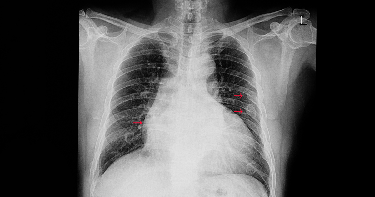
What Is a Rib Fracture?

A rib fracture is a break in one of the 12 pairs of bones forming the rib cage. Ribs protect the heart, lungs, and other vital organs. Rib fractures typically result from direct blunt trauma to the chest, such as from motor vehicle accidents, falls, or sports injuries.

Most rib fractures are uncomplicated and heal naturally within 6-8 weeks with appropriate pain management and rest. However, multiple rib fractures (flail chest) or fractures with complications can be serious and require more intensive management.

The main concern with rib fractures is not the bones themselves, but potential injuries to underlying structures. Complications can include pneumothorax (collapsed lung), hemothorax (bleeding into chest cavity), and injury to organs like the spleen, liver, or lungs.

At MyDoc Urgent Care, we provide rapid evaluation, imaging, and comprehensive management of rib fractures with emphasis on pain control and preventing serious complications.

Common Signs

Typical Symptoms

Sharp, severe chest wall pain

Pain worse with deep breathing

Pain worse with coughing or sneezing

Pain worse with movement or exertion

Painful respiration (dyspnea)

Tenderness over fracture site

Swelling or bruising over ribs

Shallow breathing (splinting)

Pain radiating to shoulder blade

Difficulty lying flat

Pain with twisting or bending

Audible cracking sensation (crepitus)

Anatomy

Rib Anatomy & Fracture Location

Different ribs have different fracture patterns and complications based on their location and surrounding anatomy.

1

Ribs 1-3 (Upper Ribs)

Location: Top of rib cage; protected by shoulder girdle

Prevalence: Less commonly fractured; requires significant force

Pain Severity: Moderate; often referred to shoulder/neck

Complications: Risk of brachial plexus, subclavian vessel injury

Healing Time: 8-12 weeks typical

Management Approach: Usually conservative; monitor for vascular injury

2

Ribs 4-8 (Mid-Ribs)

Location: Most common fracture location; most exposed

Prevalence: Most frequently fractured; common in trauma

Pain Severity: Severe; most painful to breathe

Complications: Highest risk for flail chest if multiple fractures

Healing Time: 6-8 weeks typical

Management Approach: Pain control; breathing exercises; monitor for complications

3

Ribs 9-12 (Lower Ribs)

Location: Lower rib cage; more mobile

Prevalence: Common; especially ribs 10-12

Pain Severity: Severe; pain with breathing and movement

Complications: Risk of spleen, liver, kidney injury; hemothorax/pneumothorax

Healing Time: 6-8 weeks typical

Management Approach: Conservative care; close monitoring for organ injury

4

Floating Ribs (11-12)

Location: Lower ribs not attached to sternum anteriorly

Prevalence: Less common; require significant force to fracture

Pain Severity: Variable; depends on mechanism

Complications: Can cause abdominal organ injury; may not affect breathing as much

Healing Time: 6-8 weeks typical

Management Approach: Conservative; monitor abdominal organs

Causes

Causes & Mechanisms of Rib Fractures

Rib fractures result from various causes, ranging from high-energy trauma to pathologic causes in weakened bones.

1

Blunt Trauma/Direct Impact

Description: Direct blow to chest from object, person, or surface

Prevalence: Most common cause; accounts for ~60-70% of fractures

Mechanism: Direct force causes bone to break at impact site

Severity: Variable; depends on force magnitude

Complications: Flail chest if multiple ribs broken; organ injury possible

Management: Conservative care; assess for complications

2

Motor Vehicle Accident (MVA)

Description: Trauma from motor vehicle crash or collision

Prevalence: Leading cause of rib fractures in trauma centers

Mechanism: Blunt chest trauma from steering wheel, dashboard, or seatbelt

Severity: Often severe; frequently multiple fractures

Complications: High risk of pneumothorax, hemothorax, pulmonary contusion

Management: Urgent evaluation; monitor for complications; may need admission

3

Fall from Height

Description: Trauma from falling from elevated position

Prevalence: Common cause; especially in elderly

Mechanism: Blunt impact to chest on landing

Severity: Variable; greater height = more severe

Complications: Pneumothorax, hemothorax, pulmonary injury common

Management: Imaging assessment; close monitoring

4

Blunt Chest Trauma (Sports/Recreation)

Description: Trauma from sports or recreational activities

Prevalence: Common in contact sports; accounts for ~10-15% of rib fractures

Mechanism: Direct impact during athletic competition or activity

Severity: Variable; ranges from mild to severe

Complications: Less likely to have serious complications; mainly pain

Management: Conservative care; pain management; return-to-play considerations

5

Cardiopulmonary Resuscitation (CPR)

Description: Rib fracture during chest compression for CPR

Prevalence: Occurs in ~30% of successful CPR; more with forceful compression

Mechanism: Excessive or improper chest compression force breaks ribs

Severity: Variable; usually mild to moderate

Complications: Rarely causes serious complications; considered acceptable outcome

Management: Usually self-limiting; simple pain management

6

Pathologic Fracture (Osteoporosis)

Description: Rib fracture from weakened bone without significant trauma

Prevalence: Uncommon; but increasing with aging population; ~1-3% of rib fractures

Mechanism: Bone weakened by osteoporosis breaks from minor trauma

Severity: Usually mild but concerning for underlying bone disease

Complications: Indicates need for osteoporosis assessment and treatment

Management: Conservative care; osteoporosis evaluation; bone-protective therapy

7

Pathologic Fracture (Metastatic Cancer)

Description: Rib fracture from bone weakened by cancer metastasis

Prevalence: Rare in general population; common in cancer patients

Mechanism: Cancer destroys bone structure; minor trauma causes fracture

Severity: Moderate to severe; indicates advanced cancer

Complications: Multiple complications possible; underlying cancer treatment needed

Management: Urgent oncology evaluation; may need surgical stabilization

8

Pathologic Fracture (Multiple Myeloma)

Description: Rib fracture from bone weakened by multiple myeloma

Prevalence: Common in multiple myeloma patients

Mechanism: Plasma cell infiltration destroys bone structure

Severity: Variable; but concerning for disease progression

Complications: Pain; multiple fractures; hypercalcemia

Management: Oncology-directed; may need multiple interventions

9

Severe Coughing (Tussive Fracture)

Description: Rib fracture caused by severe, prolonged coughing

Prevalence: Rare; occurs mainly in elderly or those with weakened bones

Mechanism: Forceful muscle contraction during coughing breaks bone

Severity: Usually mild to moderate

Complications: Usually isolated; rarely serious

Management: Conservative; treat underlying cough condition

10

Seizure

Description: Rib fracture from violent muscle contractions during seizure

Prevalence: Rare; occurs in ~0.1-1% of seizures

Mechanism: Forceful muscle contraction during seizure breaks bone

Severity: Usually mild to moderate

Complications: Rarely serious; mainly pain

Management: Conservative care; seizure management

Diagnosis

How Rib Fractures Are Diagnosed

Rib fracture diagnosis combines clinical examination with imaging studies. X-rays are initial imaging, but CT is more sensitive for detecting fractures and complications.

Clinical Examination

Indication: Initial evaluation of suspected rib fracture

Accuracy: 70-80% sensitivity for detecting fractures by palpation alone

Timing: Immediate; performed at bedside

Advantages: Non-invasive, immediate, no radiation, low cost

Limitations: Cannot definitively diagnose without imaging; limited in obese patients

Chest X-Ray (CXR)

Indication: Initial imaging for rib fracture; assess for pneumothorax/hemothorax

Accuracy: 60-80% sensitivity for rib fractures; misses ~20-40% of fractures

Timing: 1-5 minutes for imaging; results immediate

Advantages: Rapid, readily available, detects pneumothorax/hemothorax, low radiation

Limitations: Misses many rib fractures; cannot detect soft tissue injury

CT Chest

Indication: Suspected serious injury; assess for flail chest, organ injury, pneumothorax

Accuracy: 95%+ sensitivity for rib fractures; detects soft tissue injury

Timing: 10-15 minutes for imaging; results in minutes

Advantages: Excellent fracture visualization; detects soft tissue/organ injury; pneumothorax detection

Limitations: Higher radiation; expensive; may be overkill for uncomplicated fractures

Bone Scan (Technetium-99m)

Indication: Suspected multiple or occult fractures; stress fractures

Accuracy: 90%+ sensitivity; good for detecting multiple occult fractures

Timing: Requires 2-4 hours; delayed imaging

Advantages: Detects multiple occult fractures; good for stress fractures

Limitations: Delayed results; poor anatomic detail; not first-line for acute fractures

MRI

Indication: Suspected soft tissue injury; pulmonary contusion; detailed anatomy needed

Accuracy: 95%+ for detecting fractures and soft tissue injury

Timing: 30-60 minutes for imaging; results in hours

Advantages: Excellent soft tissue visualization; detects pulmonary contusion; no radiation

Limitations: Slower; expensive; not always necessary for simple fractures

Ultrasound

Indication: Bedside assessment of rib fractures in emergency setting

Accuracy: 80-95% sensitivity with experienced operator

Timing: 5-10 minutes; real-time results

Advantages: Rapid, portable, no radiation, bedside capable, no contrast needed

Limitations: Operator-dependent; limited by body habitus and training

Complications

Potential Complications

While most simple rib fractures are uncomplicated, serious complications can occur. Early recognition is critical.

Pneumothorax (Collapsed Lung)

Risk Profile: Occurs in 5-12% of rib fractures; higher with sharp bone fragments

Presentation: Acute dyspnea, chest pain, tachycardia, decreased breath sounds unilaterally

Timeline: Usually within first 24 hours; can occur days later

Detection: CXR or CT imaging; clinical suspicion from hypoxia/dyspnea

Treatment: Small: observation; Large/symptomatic: chest tube insertion

Hemothorax (Blood in Pleural Space)

Risk Profile: Occurs in 5-15% of rib fractures; especially lower ribs

Presentation: Dyspnea, chest pain, hypotension if significant bleeding, dullness to percussion

Timeline: Usually within first 24 hours

Detection: CXR (layering fluid) or CT imaging

Treatment: Small: observation; Large/ongoing: chest tube, possible VATS or thoracotomy

Flail Chest

Risk Profile: Occurs when 3+ consecutive ribs fractured in 2+ locations

Presentation: Severe pain, dyspnea, ineffective breathing, hypoxia, paradoxical chest wall motion

Timeline: Apparent within first 24-48 hours as swelling develops

Detection: Clinical exam (paradoxical motion); imaging (multiple fractures)

Treatment: Aggressive pain control, breathing exercises, possible stabilization or strapping

Pulmonary Contusion

Risk Profile: Occurs in 20-30% of significant rib fractures

Presentation: Dyspnea, hemoptysis, hypoxia, fever, cough, chest discomfort

Timeline: Apparent in first 24-48 hours; can worsen over days

Detection: CXR (infiltrates) or CT (consolidation); clinical assessment

Treatment: Oxygen therapy, pain control, breathing exercises, monitor for ARDS

Spleen Injury (Lower Left Ribs)

Risk Profile: Occurs with fractures of ribs 9-12 on left; present in ~5% of cases

Presentation: Left-sided chest pain, left shoulder pain (Kehr sign), hypotension, abdominal pain

Timeline: May present acutely or be delayed if contained initially

Detection: CT abdomen with IV contrast; ultrasound

Treatment: Conservative for minor injuries; splenic artery embolization or splenectomy for major

Liver Injury (Lower Right Ribs)

Risk Profile: Occurs with fractures of ribs 9-12 on right; present in ~5% of cases

Presentation: Right-sided chest pain, right upper quadrant pain, hypotension, abdominal pain

Timeline: May present acutely or be delayed

Detection: CT abdomen with IV contrast; ultrasound

Treatment: Conservative for minor; hepatic artery embolization or surgical repair for major

Kidney Injury (Posterior Ribs 11-12)

Risk Profile: Occurs with posterior rib fractures; rare; present in <2% of cases

Presentation: Flank pain, hematuria, hypotension if bleeding significant

Timeline: May be delayed if contained

Detection: CT abdomen with IV contrast; urinalysis

Treatment: Conservative for minor; surgery only if major hemorrhage or collecting system injury

Aortic Injury

Risk Profile: Rare with rib fractures alone; highest with first/second rib fractures

Presentation: Severe chest/back pain, hypotension, widened mediastinum on imaging

Timeline: May be acute or delayed rupture

Detection: CXR (widened mediastinum), CT angiography, transesophageal echo

Treatment: Urgent surgical repair

Brachial Plexus/Subclavian Vessel Injury (Ribs 1-3)

Risk Profile: Occurs with high rib fractures; rare; <1% of rib fractures

Presentation: Arm pain, paresthesias, weakness, arm swelling, diminished pulses

Timeline: Apparent immediately or shortly after injury

Detection: Clinical exam, vascular imaging (angiography, CTA, MRA)

Treatment: Depends on injury; vascular surgery for vessel injury; nerve evaluation/repair

Acute Respiratory Distress Syndrome (ARDS)

Risk Profile: Occurs with multiple severe rib fractures, flail chest, and significant pulmonary contusion

Presentation: Progressive dyspnea, hypoxia unresponsive to O2, tachypnea, bilateral infiltrates

Timeline: Usually within 24-72 hours after injury

Detection: CXR (bilateral infiltrates), clinical assessment, blood gas analysis

Treatment: ICU admission, mechanical ventilation, lung protective strategies

Treatment

Management of Rib Fractures

Most rib fractures are managed conservatively with pain control, rest, and gradual return to activity. Healing typically takes 6-8 weeks. Proper pain management is critical to allow breathing exercises and prevent pneumonia.

Acute Phase (First 24-48 Hours)

Goals: Stabilize patient, assess for complications, initiate pain control

Key Interventions:

  • Immediate imaging (CXR, CT if indicated) to assess for pneumothorax/hemothorax
  • High-flow oxygen if hypoxic or suspected pulmonary contusion
  • IV pain control (opioids if severe; NSAIDs contraindicated initially)
  • Pulse oximetry monitoring
  • Assessment for complications requiring urgent intervention
  • Bed rest initially; gradual mobilization as tolerated

Monitoring: Vital signs, oxygen saturation, respiratory status, signs of complications

Subacute Phase (3-7 Days)

Goals: Optimize pain control, promote mobilization, prevent complications

Key Interventions:

  • Transition to oral pain medications (NSAIDs now appropriate)
  • Rib belt or elastic binder if needed for comfort (controversial)
  • Breathing exercises (splinting); incentive spirometry if available
  • Gradual increase in activity as tolerated
  • Physical therapy for pain management and mobility
  • Repeat imaging only if clinical deterioration

Monitoring: Pain control, respiratory function, signs of complications, functional improvement

Recovery Phase (1-8 Weeks)

Goals: Progress rehabilitation, return to normal activities gradually

Key Interventions:

  • Continue pain management (usually decreased need by 2-4 weeks)
  • Progressive increase in activity; avoid heavy lifting/contact sports
  • Breathing exercises continue; graduated physical therapy
  • Return to work as tolerated (may need job modification initially)
  • Graduated return to sports (typically 6-8 weeks for simple fractures)
  • Home exercises for strength and flexibility

Monitoring: Functional recovery, pain trends, complications, return-to-activity readiness

Long-term (2-3 Months+)

Goals: Full functional recovery and return to preinjury activities

Key Interventions:

  • Most pain resolved by 6-8 weeks; persistent pain rare
  • Full return to activity including contact sports
  • Continue strength/flexibility exercises
  • Address any chronic pain or functional limitations
  • Reassurance that ongoing mild discomfort can persist for months

Monitoring: Resolution of symptoms, functional status, late complications (post-traumatic arthritis rare)

Pain Control

Pain Management Options

Effective pain control is essential for rib fracture recovery. Adequate analgesia allows breathing exercises and prevents pneumonia.

Nonsteroidal Anti-Inflammatory Drugs (NSAIDs)

Indication: First-line for pain management after acute phase (48+ hours)

Effectiveness: Excellent pain relief; 70-80% of patients satisfied

Onset: 30-60 minutes for effect

Advantages: Effective, oral administration, inexpensive, reduces inflammation

Disadvantages: GI upset, ulcer risk, renal effects, avoid if hypotension

Acetaminophen

Indication: Alternative analgesic if NSAIDs contraindicated

Effectiveness: Moderate pain relief; 50-60% of patients satisfied

Onset: 30-60 minutes

Advantages: Oral, safe, well-tolerated, no GI effects

Disadvantages: Less effective than NSAIDs, hepatotoxicity if overdosed

Opioid Analgesics

Indication: Severe pain uncontrolled by NSAIDs/acetaminophen; first 24-48 hours

Effectiveness: Excellent pain relief but risk of dependence

Onset: 15-30 minutes oral; 5-15 minutes IV

Advantages: Excellent pain control, IV option for severe pain

Disadvantages: Respiratory depression risk, addiction/dependence risk, constipation, nausea

Local Nerve Blocks

Indication: Severe localized pain; alternative to systemic opioids

Effectiveness: Excellent localized pain relief; 80-90%

Onset: 15-30 minutes

Advantages: Excellent pain control, preserves respiratory function better than opioids

Disadvantages: Procedural risks, requires specialist, not universally available

Rib Binder/Elastic Tape

Indication: Pain management and splinting; controversial

Effectiveness: Moderate pain relief; 50-70% satisfaction

Onset:

Advantages: Non-pharmacologic, inexpensive, can facilitate activity

Disadvantages: May restrict breathing; increases pneumonia risk; less recommended now

Ice/Cold Compression

Indication: First 24-48 hours for pain and swelling

Effectiveness: Moderate pain relief; adjunctive benefit

Onset: 10-20 minutes

Advantages: Non-pharmacologic, safe, inexpensive, reduces swelling

Disadvantages: Short duration, need frequent reapplication

Heat Therapy

Indication: After 48-72 hours; may help muscle tension

Effectiveness: Mild to moderate relief; mainly for muscle tension

Onset: 10-20 minutes

Advantages: Non-pharmacologic, safe, inexpensive

Disadvantages: Less evidence than cold initially; can increase swelling if used too early

Physical Therapy/Breathing Exercises

Indication: Adjunctive pain management; starts within days

Effectiveness: Moderate to excellent; 70-85% benefit

Onset: Days to weeks

Advantages: Reduces opioid need, prevents complications, improves outcomes

Disadvantages: Requires patient compliance, not universally available

Pain Control is Critical: Uncontrolled pain leads to splinting (shallow breathing), which increases pneumonia risk and delays healing.

Take Pain Medications Regularly: Don't wait until pain is severe. Regular dosing maintains better control and improves outcomes.

Prevention

Preventing Rib Fractures

While not all rib fractures can be prevented, many can be avoided through safety precautions and bone health.

Always wear seatbelt while driving; position correctly across lap/shoulder

Use airbags in vehicles; ensure properly maintained

Practice safe driving; avoid excessive speed and reckless driving

Wear protective equipment during contact sports (chest protectors available)

Ensure home safety; remove tripping hazards, adequate lighting, grab bars

Prevent falls in elderly: strength training, balance exercises, vision correction

Avoid climbing ladders/heights without proper safety equipment

Use proper body mechanics when lifting; avoid heavy lifting

Maintain bone health: adequate calcium, vitamin D, weight-bearing exercise

Avoid smoking; smoking decreases bone density

Limit alcohol consumption; excess alcohol impairs bone health

Regular exercise maintains muscle strength and bone density

Address osteoporosis with treatment if diagnosed

Fall prevention in elderly through home modifications and medical management

CPR training uses correct technique with appropriate force

Self Care

Care at Home

Proper home care accelerates healing and prevents complications. Follow these guidelines for optimal recovery.

Rest initially; gradually increase activity as tolerated and pain permits

Take pain medications regularly (not just when pain severe) for better control

Apply ice for first 24-48 hours; then heat may help

Perform breathing exercises regularly (splinting); use pillow to support ribs

Use incentive spirometer if available; helps prevent pneumonia

Sleep in semi-recumbent position (propped up); fully supine more painful

Avoid heavy lifting and strenuous activity for 4-6 weeks minimum

Avoid contact sports for 6-8 weeks or until pain-free

Practice good posture; don't collapse forward, causes more pain

Avoid coughing fits; treat cough with medications if needed

Avoid sudden movements; move slowly and deliberately

Support ribs with pillow when coughing or sneezing

Wear loose-fitting clothing; avoid constrictive garments

Stay hydrated; proper hydration aids healing

Eat nutritious diet with adequate protein and calcium

Alert

When to Seek Immediate Care

Certain symptoms require immediate emergency evaluation. Do not wait if you experience these warning signs.

Severe Shortness of Breath

Sudden or worsening dyspnea suggests pneumothorax or hemothorax

Coughing Blood

Hemoptysis suggests pulmonary contusion or massive bleeding

Severe Chest Pain

Pain that worsens despite pain medications or new onset severe pain

Fever >101°F

Fever suggests infection/pneumonia; risk with rib fractures

Cyanosis or Severe Hypoxia

Blue lips/fingers or persistent low oxygen despite treatment

Collapsed Chest Wall

Paradoxical inward motion of chest wall indicates flail chest

Abdominal Pain/Distention

Suggests splenic, liver, or other abdominal organ injury

Decreased Breath Sounds One Side

Suggests pneumothorax or hemothorax requiring urgent intervention

Confusion or Altered Mental Status

Suggests severe hypoxia or respiratory failure

Unable to Breathe Lying Down

Severe orthopnea suggests pulmonary contusion or other complication

Most Rib Fractures Heal Well: With proper pain management and care, most uncomplicated rib fractures heal completely within 6-8 weeks with no long-term consequences.

Complications Can Occur: While rare, serious complications like pneumothorax or organ injury require urgent diagnosis and treatment. Early recognition is critical.

Pain Control is Essential: Adequate analgesia allows breathing exercises, prevents pneumonia, and speeds recovery. Don't suffer through pain.

At MyDoc Urgent Care, we provide rapid evaluation, imaging, and comprehensive management of rib fractures. We assess for complications, provide effective pain control, and develop recovery plans tailored to your needs. Visit one of our convenient locations in Forest Hills, East Meadow, Brooklyn, Bronx, Jackson Heights, or Little Neck for expert trauma evaluation and care.